Dr. Starcevic is specialized in evaluation, diagnosing and treatment of cardiovascular disease. After being evaluated by the doctor with detailed and personalized history taking and thorough physical exam a patient might require testing to delineate cardiac structure and function. Here is more information…
common cardiovascular tests
Your doctor can order and perform different tests to see if your symptoms are caused by underlying cardiac or vascular conditions. Here is a list of some common tests either performed in the office or at an outpatient facility:
- Ankle-brachial index (ABI): A noninvasive exam that compares the blood pressure in your feet to the blood pressure in your arms to determine how well your blood is flowing.
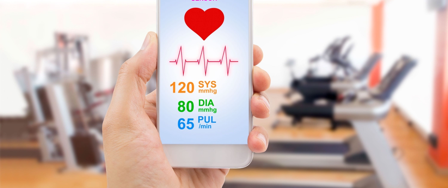
- Blood Pressure: During this commonly performed office test, the cuff is placed around the upper arm before being manually or electronically inflated and then slowly deflated to determine if a person has high blood pressure.
- Blood tests: Ordered to diagnose or rule out a medical condition and assess response to drug therapy.
- Cardiac Catheterization: Examines the inside of your heart’s blood vessels using special X-rays called angiograms; a contrast agent is injected into the artery in your arm or groin through a thin flexible tube and X-rays are taken to show blood flow; it gives information about blockages in coronary arteries, function of the heart valves, heart muscle and pressures in the heart.
- Cardiac Computed Tomography: Computer imaging (tomography) refers to several non-invasive diagnostic-imaging tests that use computer-aided techniques to gather images of the heart. A computer creates three-dimensional (3-D) images that can show blockages caused by calcium deposits you may have in your coronary arteries.
- Cardiac MRI: Noninvasive imaging test that uses radio waves, magnets, and a computer to create detailed pictures of your heart.
- Cardioversion: A procedure used to return an irregular heart rhythm to normal through an electric shock or drug; it is performed in a closely monitored hospital-based setting, such as an intensive care unit, an emergency department, or a specially equipped procedure room. The patient’s heart rate and rhythm, blood pressure, breathing rate, and oxygen levels are monitored.
- Chest X-Ray: Takes a picture of the heart, lungs and bones of the chest. Determines whether the heart is enlarged or if fluid is accumulating in the lungs as a result of the heart attack or heart failure.
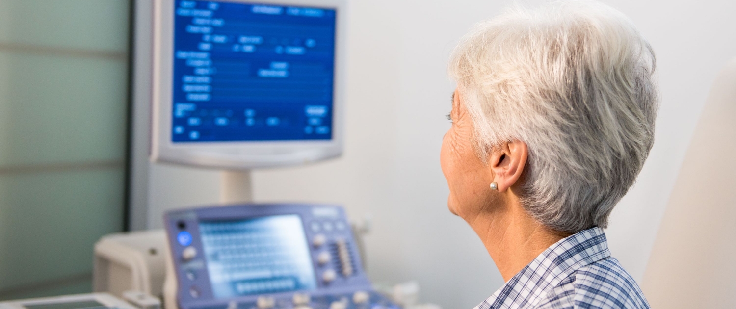
- Echocardiogram: A hand-held device placed on the chest that uses high-frequency sound waves (ultrasound) to produce images of your heart’s size, structure and motion, provides valuable information about the health of your heart.
- EKGs (Electrocardiogram): Records the electrical activity of the heart including the timing and duration of each electrical phase in your heartbeat.
- Electrophysiology Studies (EPS): Tests that help doctors understand the nature of abnormal heart rhythms and to find where an arrhythmia (abnormal heartbeat) is coming from. During EPS, doctors insert a thin tube called a catheter into a blood vessel that leads to your heart. A specialized electrode catheter designed for EP studies lets them send electrical signals to your heart and record its electrical activity.
- Event Recorder: A monitoring device which is attached to the patient’s chest to detect irregularities in heartbeat. Event monitoring is a way to record the heart rhythm when your symptoms occur less than once a day and can be worn up to for up to 30 days.
- Exercise Stress Test: The most common kind of cardiac stress test; electrodes that are attached to the skin on the chest area monitor and record your heart function while you walk in place on a treadmill. Many aspects of your heart function can be checked including heart rate, breathing, blood pressure, ECG (EKG) and how tired you become when exercising.
- Holter Monitor: A small recording device which is attached to the patient’s chest to document the electrical activity of the heart during daily activities to help doctors determine the condition of the heart, usually worn for 24 hours.
- Implantable Loop Recorder: A device that can record your heart rhythm for up to 36 months. This device is placed under the skin through a minor operation. This is the best way to record very serious rhythm problems that may be happening only rarely.
- Nuclear Stress Test: Is a non-invasive imaging test that shows how well blood flows through (perfuses) your heart muscle. It can show areas of the heart muscle damage or reduced blood flow. This test is often called Myocardial Perfusion Imaging:
- It is similar to a routine exercise stress test but with images of the heart muscle created with a special camera when a small amount of radioactive substance is injected into the bloodstream when patient is at maximum level of exercise.
- Pacemaker Interrogation: A test on a device implanted in the chest to regulate the heartbeat.
- Tilt-Table Test: Doctors use tilt-table tests to find out why people feel faint, are lightheaded or completely pass out. During the test, you lie on a table that is slowly tilted upward. The test measures how your blood pressure and heart rate respond to the force of gravity while your blood pressure and heart rate are closely monitored.
- Transesophageal Echocardiography (TEE): Uses high-frequency sound waves (ultrasound) to make detailed pictures of your heart on a video screen; Doctors often use TEE when they need more details about your heart than a standard echocardiogram can give them. The echo transducer for a TEE is attached to a thin tube that passes through your mouth, down your throat and into your esophagus.
- Ultrasound Imaging: A non-invasive method that visualizes the arteries and veins with the use of high frequency sound waves and measures the blood flow to indicate the presence of a blockage or disease in the area of interest.

